Additional EM structure validation now available through OneDep
The wwPDB validation reports provided in the OneDep system now have additional validation for electron microscopy (EM) maps to help users identify potential discrepancies in their data.
EMDataResource played a key role in development of these new features for structures deposited to EMDB and PDB.1,2
The updated wwPDB validation reports in the OneDep system now incorporate an extensive EM map validation process, integrating a range of established validation methods for EM data previously available on the EMDB pages. Initially, this additional EM validation is only provided to depositors in the OneDep system, however in future will be provided for entries throughout the PDB and EMDB archives.
The process includes an analysis of the fit of the PDB model to the EMDB map, represented at an amino acid level on the residue-property plots and globally by a visual overlay of the map and model (see below images). FSC curves are also included to compare reported and estimated resolution, where either half maps or FSC data was uploaded.
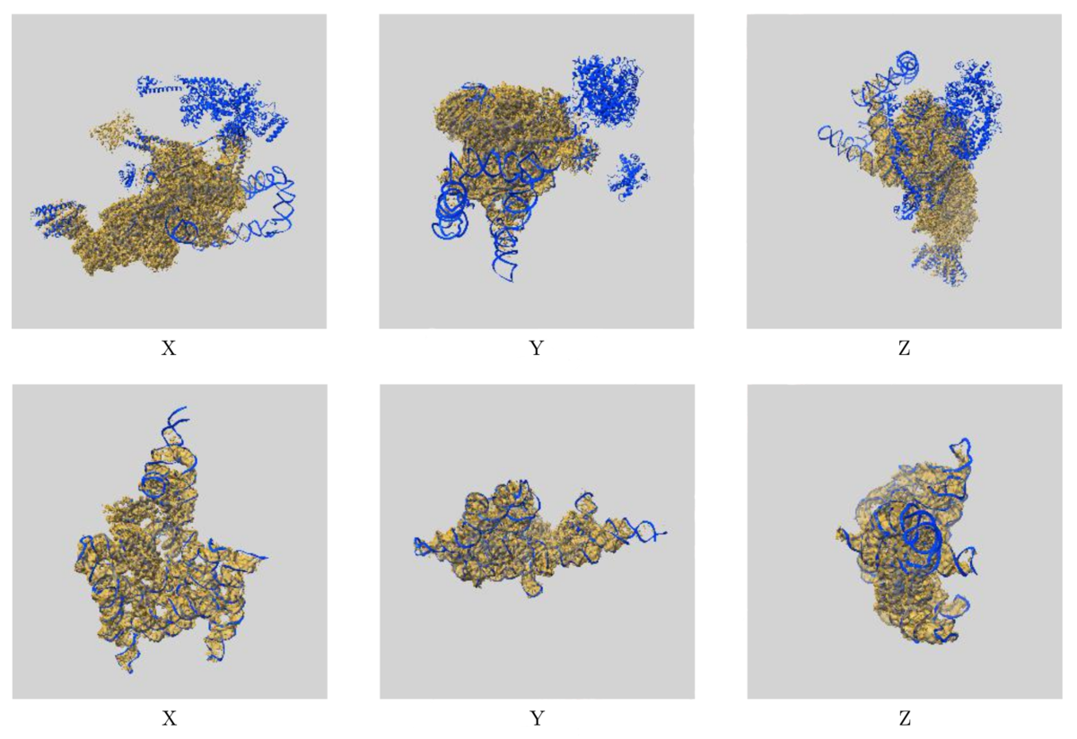
Images showing the visual EM map/model fit as displayed in the new validation reports. The top images display three orthogonal views of the map and model for EMD-0360 and 6N7P and highlights where there are regions of the model not covered by the EM map at the provided contour level, while the below images show the fit of EMD-9105 and 6ME0, where the majority of the model fits well to the map at the provided contour level.
The additional EM validation also includes various graphics for visual inspection of the data. Included in the reports are images of orthogonal projections, central slices, mask visualisation, and more, allowing for inspection of details in the map and identification of artifacts. A statistical analysis of the EM map volume is also provided, including graphs of map density distribution, volume estimate by contour, and rotationally averaged power spectrum, providing more thorough analysis of the EM volume.
These changes should help both depositors and users to identify potential errors in EM data and give more clarity about potential limitations of the data in both the PDB and EMDB.
Additional information about validation reports for EM entries is available here.
For questions or queries about wwPDB validation, please contact validation@mail.wwpdb.org.
Source: wwPDB News
References
- Web-based visualisation and analysis of 3D electron-microscopy data from EMDB and PDB. Lagerstedt I, Moore WJ, Patwardhan A, Sanz-García E, Best C, Swedlow JR, Kleywegt GJ. J Struct Biol. 2013 Nov;184(2):173-81. doi
- Abbott S, Iudin A, Korir PK, Somasundharam S, Patwardhan A. EMDB Web Resources. Curr Protoc Bioinformatics. 2018 Mar;61(1):5.10.1-5.10.12. doi